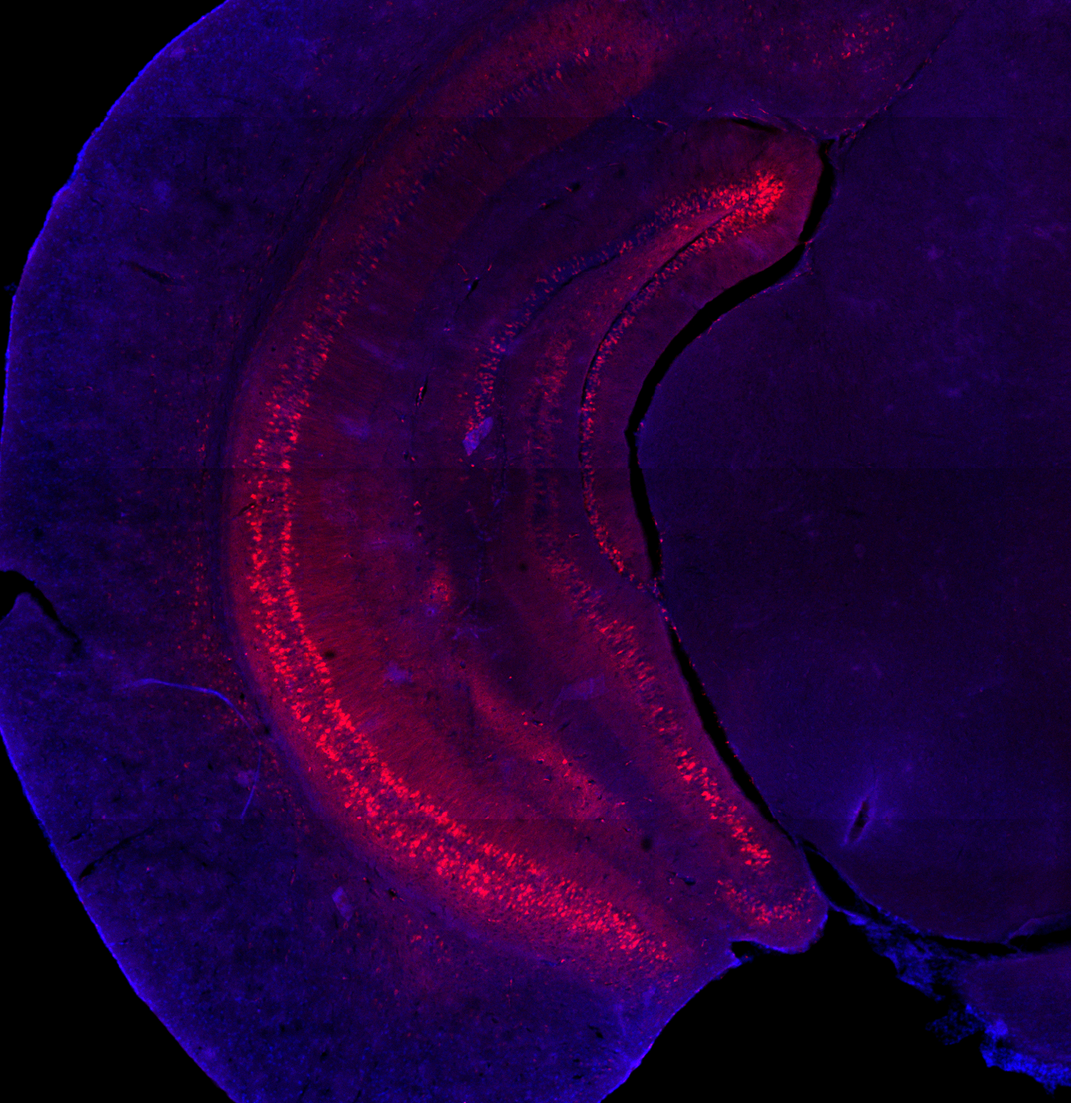
VHPC-PFC Projections
Pyramidal cells of the ventral hippocampus (VHPC) send axonal projections to the prefrontal cortex (PFC), and these direct inputs critically support the encoding of cues during spatial working memory.
A mouse undergoing the open-field social interaction test. The animal’s body position and orientation are tracked by a neural network trained using DeepLabCut machine vision software.
A mouse performing the 4-goal version of the t-maze delayed non-match-to-sample spatial working memory task during optogenetic manipulation of VHPC-PFC axon terminals.

PFC place coding
A topological heat map of activity from an example PFC neuron during the spatial working memory task. Warmer colors and higher elevation signify elevated firing rate at a given point in the 2-dimensional environment.
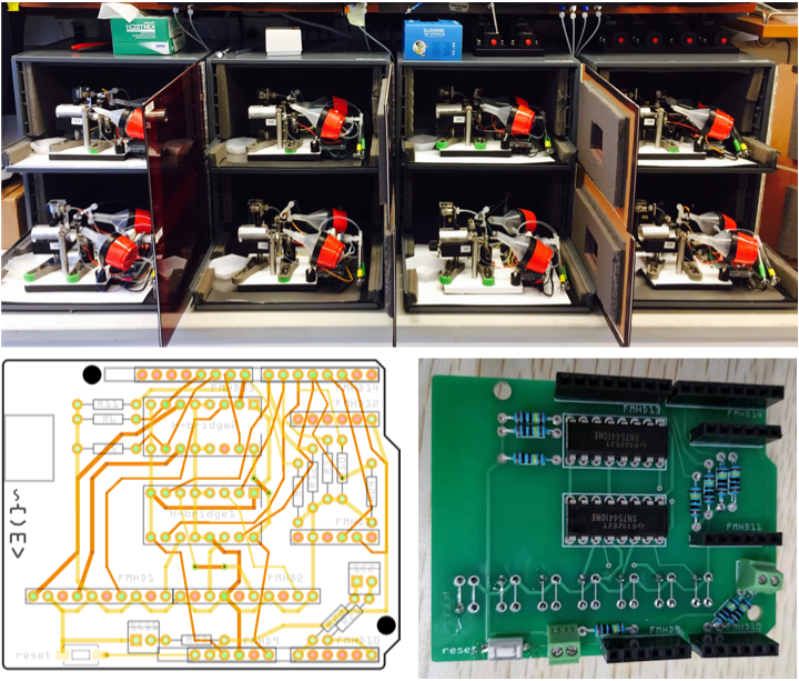
set-shifting task
Custom-designed behavioral rigs, powered by custom-printed circuit boards, for high-throughput behavioral testing of serial extra-dimensional set-shifting. Mice receive odor and whisker cues on each trial and learn, through trial and error, which sensory modality to attend to for a reward.
Video of calcium activity from spontaneous firing of 953 identified PFC neurons in prelimbic (upper field) and infralimbic (lower field) areas.
Trial-averaged activity for each of 953 individually-identified neurons during the set-shifting task.
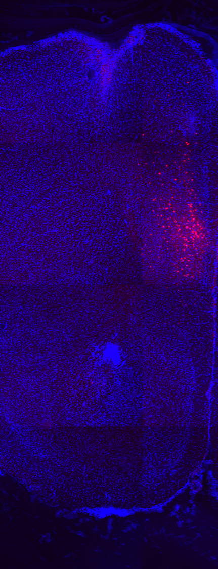
PFC-VMS Cell bodies
Pyramidal cells in layers 2/3 and 5 of the prefrontal cortex send axonal projections to the ventromedial striatum (VMS). These neurons, as well as those projecting to the medial dorsal thalamus, are critically important for encoding feedback information about the outcome of each trial in order to guide responses on future trials.
A composite video of calcium activity (green) in PFC neurons, and specifically in PFC-VMS neurons (green + red).
Light field microscopy allows for the simultaneous capture of tissue volumes, rather than 2D planes. Here a volume of fluorescent beads is reconstructed from a light field image and compared with a 2-photon z-stack.
Volumetric video of spontaneous calcium activity in PFC, captured by light field imaging and reconstructed from SWIFT reconstruction algorithm (Grosenick et al, 2017). Lower right: XY view; upper right: XZ view; lower left: YZ view. Frame rateL 80Hz